- Browse Category
Subjects
 We Begin at the EndLearn More
 - Choice Picks
- Top 100 Free Books
- Blog
- Recently Added
- Submit your eBook
- Browse Category
We Begin at the EndLearn More
- Choice Picks
- Top 100 Free Books
- Blog
- Recently Added
- Submit your eBook

In Situ Raman Microscopy Of A Single Graphite Microflake Electrode In A Li(+)-containing Electrolyte
2021-01-05 00:14:11
Highly detailed Raman spectra from a single KS-44 graphite microflake electrode as a function of the applied potential have been collected in situ using a Raman microscope and a sealed spectroelectrochemical cell isolated from the laboratory environ...
Read more
Highly detailed Raman spectra from a single KS-44 graphite microflake electrode as a function of the applied potential have been collected in situ using a Raman microscope and a sealed spectroelectrochemical cell isolated from the laboratory environment. Correlations were found between the Raman spectral features and the various Li(+) intercalation stages while recording in real time Raman spectra during a linear potential scan from 0.7 down ca. 0.0V vs Li/Li(+) at a rate of 0.1 mV/s in a 1M LiClO4 solution in a 1: l (by volume) ethylene carbonate (EC): diethyl carbonate (DEC) mixture. In particular, clearly defined isosbestic points were observed for data collected in the potential range where the transition between dilute phase 1 and phase 4 of lithiated graphite is known to occur, i.e. 0.157 Less

Compare Prices
| Store | Availability | Book Format | Condition | Price |
|---|---|---|---|---|
| Indigo Books & Music | In Stock | Paperback | Paperback | Buy CAD 16.99 |
Available Discount
No Discount available
Related Books
View All
Made for Me

The Silent Woman (Cat Carlisle, Book 1)

American Moonshot: John F. Kennedy and the Great Space Race
Diary of an Awesome Friendly Kid: Rowley Jefferson's Journal
Can't Make This Stuff Up!: Finding the Upside to Life's Downs

We Are Not Friends (You Are Not Small)
The Wonderful Things You Will Be
Secrets Between Us: An absolutely gripping psychological thriller

Life Will Be the Death of Me: ...And You Too!

Commander in Cheat: How Golf Explains Trump

Good Night, Little Blue Truck
Hurricane Season

You Be You: Detox Your Life, Crush Your Limitations, and Own Your Awesome

House of Shadows: An Enthralling Historical Mystery

Going to School

Feed Your Best Friend Better: Easy, Nutritious Meals and Treats for Dogs

There Was an Old Lady Who Swallowed a Chick!

Twinkle, Twinkle, Little Star (Indestructibles Series)
Clean & Lean: 30 Days, 30 Foods, a New You!

Trillion Dollar Coach: The Leadership Playbook of Silicon Valley's Bill Campbell

The Book Club
Falter: Has the Human Game Begun to Play Itself Out?

Row, Row, Row Your Boat (Indestructibles Series)

The Secret Wife: A captivating story of romance, passion and mystery
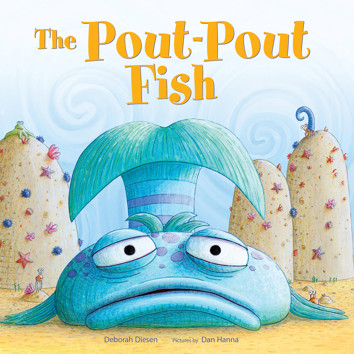
The Pout-Pout Fish

Settings
Reflow text when sidebars are open.